
Two parts comprise the hip joint: a ball on the upper end of the thigh bone (femur) called the femoral head, and a socket in the pelvis known as the acetabulum. In a normal hip, the head of the femur rotates freely within the smooth, concentric surface of the acetabulum. A smooth, low-friction coverage of “hyaline” cartilage lines the joint, and joint fluid lubricates the articulation to allow smooth, frictionless, smooth motion. The acetabulum is lined by the labrum, which is a highly innervated fibrocartilaginous structure. The labrum acts as a gasket seal between the femoral head and the acetabulum and adds to the joint stability by deepening the socket. The articulation is encapsulated by a joint capsule and ligamentous complex that further stabilizes the hip articulation. Injury and degeneration of any of these structures can cause hip pain sufficient for a patient to seek medical attention.
The Normal Hip: Osseous Anatomy
The normal acetabulum “covers” the upper (superior) portion of the head of the femur as well as a partial portion of the front (anterior) and back (posterior) femoral head. The femoral head is oriented (angled) into the acetabulum. This relationship allows the force of weight-bearing to be distributed throughout the acetabular cartilage surface, and allows stable motion due to the containment of the head within the socket. Insufficient coverage of the head by the acetabulum, as in hip dysplasia can lead to instability and abnormally high stress on the soft tissues (cartilage and labrum) in the periphery of the socket.
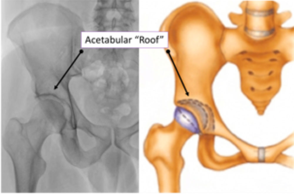
Figure: The Hip Joint
A normal relationship between the acetabulum (socket) and the femur (ball). The acetabulum covers the femoral head to keep it “contained” and stable during weight bearing and motion. The femur is oriented into the socket, and force is distributed throughout the cartilage of the acetabulum.
In a normal hip develops as precisely fitting parts, allowing smooth range of motion throughout all positions of the leg. The shapes of the femoral head and of the acetabulum are specifically structured to allow stable, physiological motion without conflict, or “impingement” (FAI) of one upon the other. The ball is circular, and the socket oriented to the front (anterior) of the body to allow the leg to come up into flexion without the ball hitting the socket. If the ball is not sufficiently round (CAM impingement) or the socket excessively covers the ball (Pincer Impingement), the femur may hit the acetabular rim during motion.
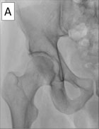
A. Normal Hip. The ball is round and fits perfectly into the socket, which is oriented anteriorly to allow conflict free motion.
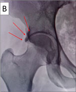
B. Pincer Impingement. There is significant “over coverage” or overhang of the socket. The femur will hit the socket early in hip flexion.
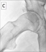
C. Cam Impingement. The femoral head is “out of round” and, without a recess like the normal hip, hits the acetabular rim with hip flexion.
Abnormalities in the osseous architecture leading to dysplasia or FAI can cause injury and degeneration of the soft tissues within the hip as well as problems with the muscles around the hip. Injury to any of these structures-the acetabular labrum, the hyaline cartilage, the ligaments and hip musculature, can cause pain and disability. At the Nashville Hip Institute at TOA, Dr. Byrd, along with his masterful staff of practitioners, provide a comprehensive evaluation and diagnostic program to identify the most likely cause of every individual’s unique hip pain.
Location
2004 Hayes Street
Suite 700
Nashville, TN 37203
Office Hours
Monday-Friday:
8:00 am – 5:00 pm